A new ultrasound technology that could pick up far more cases of cancer and cut the need for biopsies has been developed by scientists.
At present, ultrasounds can help identify potential problems with key organs but are not sensitive enough to detect cancer.
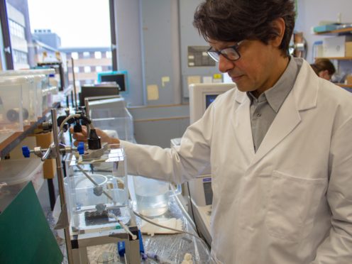
Now a team at Heriot-Watt University in Edinburgh say they have made a major breakthrough and can produce images with five to 10 times the resolution of traditional methods.
The new technique allows organs and blood flow to be scanned in super-resolution for the first time, with NHS trials on people starting at Edinburgh’s Western General Hospital in December.
The process works by injecting tiny bubbles into the bloodstream and scanning organs so that the blood flow can be shown with 0.05mm precision.
The patient need only stay still for a few minutes while they are scanned – allowing images to be produced in reasonable time.
Computer technology is able to track these bubbles to produce images which have a greater resolution than anything in use at the moment.
By looking at blood vessels and flow, experts are able to map the networks that are enabling cancerous tumours to grow.
Dr Vassilis Sboros, from Heriot-Watt University, who led the research, said: “What we can see is all these bubbles one by one – we see dots in the image.
“By joining the dots we end up with a picture that has much more detail and a lot more specific information.
“At the moment we can detect a few cancers with ultrasound but our new technique increases the confidence with which we can be sure whether something looks cancerous.
“We now need to do clinical trials on humans, but we may well be able to pick up cancers, such as pancreatic cancer and liver cancer, far earlier.”
Dr Sboros said the new technique will not require hospitals to upgrade their current equipment.
He said: “The limitations of current ultrasound images mean more expensive techniques like MRI are often employed for diagnosis and treatment.
“MRI doesn’t provide clinicians with more detail but it has generally provided better results than other methods.
“However, in the prostate for example, biopsy has to be performed as a separate procedure which is more expensive for the hospital and can be both disruptive and distressing for the patient.
“Due to the super-resolution capability of our new images, we anticipate that the ability of the medical staff to pinpoint, diagnose and treat a range of cancers will be greatly enhanced.
“We will work to establish the usefulness of our method in the upcoming clinical study.
“We hope that further research will help expand this method to other applications in cardiovascular disease, diabetes, liver disease and transplant rejection and one day biopsies may not be necessary.”
A study of the technique published in the Journal of Investigative Radiology showed that images of prostate cancer could be created.
Professor Alan McNeill, consultant urological surgeon at the Western General Hospital in Edinburgh said: “Prostate cancer is an increasing problem for our society.
“Whilst we have a number of methods for detecting it, these don’t always provide us with the important information that we need regarding who has cancer that needs to be treated and who doesn’t.
“A method that maps the blood flow of the tumour accurately could well provide new information about the disease state that allows us to better identify those men who need urgent treatment and those who don’t.
“It is exciting that we will be the first hospital in the world that will assess this method with patients.”